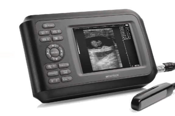
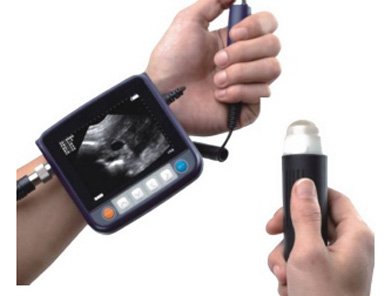
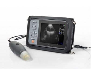
Sonovet Palm Veterinary Ultrasound Scanner with Rectal Probe
- High-quality veterinary ultrasound scanner with a 5.5 inch TFT display (Option: 7 inch)
- Features full digital beam-forming technology and probe automatic identification
- Offers a wide range of scanning methods including Convex, Micro-convex, Linear, and rectal
- Equipped with 256 frames for cine-memory and 64 image permanent storage
- SKU
- A3CBA1D90234
- Brand
- Teds Medical
- Availability
- In stock
- ✓ Worldwide shipping


Sonovet Palm Veterinary Ultrasound Scanner with Rectal Probe
Compact, portable veterinary ultrasound system featuring a 5.5-inch TFT display with full digital beam-forming technology and probe automatic identification. Delivers high-resolution imaging across multiple scanning modalities with 256-level grayscale and comprehensive measurement capabilities for equine, small animal, and obstetric applications.


Clinical overview
The Sonovet Palm is a lightweight, battery-powered veterinary ultrasound scanner engineered for field and clinical use. Its full digital beam-forming architecture delivers consistent image quality across convex, micro-convex, linear, and rectal scanning modalities, making it suitable for equine reproductive assessment, small animal abdominal imaging, and obstetric evaluation. The system supports real-time cine-loop recording (256 frames) and permanent image storage (64 images), enabling clinicians to capture and review dynamic anatomy with precision.
Automatic probe identification and multi-frequency transducers (3.5–8.5 MHz) allow rapid protocol switching without manual configuration. Comprehensive measurement packages include distance, area, circumference, cardiac parameters (heart rate, EF), and obstetric indices (GA, fetal weight, EDD), streamlining diagnostic workflow and report generation.
Key features & surgeon benefits
Full Digital Beam-Forming & Grayscale
- 256-level grayscale for superior contrast resolution
- Gamma correction and histogram adjustment for optimized image processing
- Four selectable focusing combinations and four color-coded modes
- Real-time display of focus position, gain (near, far, overall), and scanning parameters
Rapid Protocol & Data Capture
- Automatic probe identification eliminates manual setup
- 256-frame cine-memory for real-time video review and frame selection
- 64-image permanent storage with left/right, up/down, brightness, and contrast adjustment
- USB 2.0 connectivity for direct image transfer to PC and mouse interface support
Comprehensive Diagnostic Tools
- 15 body mark types with customizable calipers
- Obstetric measurements: GA, CRL, BPD, HC, FL, AC, fetal weight, EDD calculation
- Cardiac assessment: heart rate, EF rate, time-based measurements
- Automatic clinic report generation with date, time, patient ID, and physician annotation
Multi-Frequency Probe Options
- Standard: 7.5 MHz linear rectal probe (multi-frequency 7.5, 8.5 MHz)
- Optional: 3.5 MHz convex, 5.0 MHz micro-convex, 6.5 MHz transvaginal probes
- Scanning modes: B, B+B, 4B, B+M, M display options
- Image magnification: ×1.0, ×1.2, ×1.5, ×2.0 times
Technical specifications
| Indications | Equine, small animal, and obstetric/gynecological ultrasound imaging |
|---|---|
| Display | 5.5-inch TFT screen (7-inch option available) |
| Grayscale | 256 levels |
| Scanning methods | Convex, micro-convex, linear, rectal |
| Display modes | B, B+B, 4B, B+M, M |
| Cine-loop memory | ≥400 frames |
| Permanent storage | 128 frames |
| Display depth | ≥220 mm, multi-step adjustable |
| Image magnification | ×1.0, ×1.2, ×1.5, ×2.0 |
| Focusing combinations | 4 selectable options |
| Color modes | 4 different coded-color options |
| Body marks | 15 types with customizable calipers |
| Measurement capabilities | Distance, circumference/area, time/heart rate/EF, obstetric indices (GA, CRL, BPD, HC, FL, AC, fetal weight, EDD) |
| Image processing | DFS, DRF, RDA, VGA/DSC, post-processing, gamma correction, histogram |
| Connectivity | USB 2.0 (real-time image upload to PC), PAL-D video output, NTSC for video recorder |
| Operating time | ≥2 hours |
| Net weight | 0.8 kg |
| Sterilisation | As applicable |
Standard instrument composition
| Item | Description | Qty |
|---|---|---|
| Main unit | Sonovet Palm ultrasound scanner with 5.5-inch TFT display | 1 |
| Rectal probe | L60/6.5 MHz linear rectal probe (multi-frequency 7.5, 8.5 MHz) | 1 |
| USB cable | USB 2.0 for PC connectivity and mouse interface | 1 |
| Video cable | PAL-D output for video recorder and workstation connection | 1 |
Optional accessories & probes
| Item | Description | Frequency |
|---|---|---|
| Convex probe | R40 convex probe (multi-frequency) | 3.5, 5.0 MHz |
| Micro-convex probe | R20 micro-convex probe (multi-frequency) | 4.5, 5.0, 5.5 MHz |
| Linear probe | L40 linear probe (multi-frequency) | 7.5 MHz |
| Transvaginal probe | 6.5 MHz transvaginal probe (multi-frequency) | 6.5, 7.5 MHz |
| PC software | Image transfer and management software | — |
| Video printer | Thermal video printer for image documentation | — |
| Car lighter | 12V car power adapter | — |
| Ultrasound goggles | Optional viewing accessory | — |
Get in touch for pricing & availability
Contact our sales team to discuss system configuration, probe options, and pricing. We provide technical support and can arrange demonstrations for qualified veterinary clinics and equine facilities.
Email: [email protected]
Available on request
Configuration, paperwork & clinical fit
The full spec + documentation pack for this device ships ahead of purchase on request. A sales engineer responds within one business day.
Configuration sheet
Specifications, accessories & clinical fit
Sizes and dimensions, material and finish, compatible instruments and screw references, what ships in the set, lead time and warranty — all in one specification sheet, sent within one business day.
Documents & downloads
Regulatory paperwork & manuals
This device's IFU, CE Declaration of Conformity, ISO 13485 certificate and (where applicable) FDA 510(k) clearance ship with the order. Reach out below for the pack ahead of purchase.
Get in touch
Need configuration help, certificates, or volume pricing?
Our clinical sales team will scope quantities, voltage, accessories and regulatory paperwork — usually within one business day.
Customer reviews
No reviews yet — be the first to share your experience.
Related products
See all →
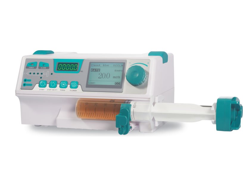
MD910 Veterinary Syringe Pump
- Audible and visual alarms for various scenarios such as occlusion, low battery, and end of infusion - Compatible with syringes of different sizes from 10ml to 60ml of any brand - Three work modes: Rate mode, Time Volume mode, Dosage Weight mode - KVO feature automatically opens to keep the vein open after infusion completion

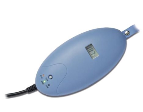
MD-T1 Veterinary Fluid Blood Warmer
- Maintain body temperature during and after anesthesia - Prevent risks such as shock, heart problems, and hypothermia - Suitable for use with standard fluids, blood, and TPN fluids - Lightweight, portable, and easy to set up - Compatible with standard tubing sets for small animals - Helps decrease temperature difference between animal and IV liquid - Ensures safe infusion of warm fluid without the need for disposables
SonovetID Veterinary Ultrasound Scanner with RFID Technology Handheld 6 Inch Color Display
- Handheld veterinary ultrasound scanner with RFID technology - 6-inch high-resolution color display for clear imaging - Level 7 waterproof design for durability - Built-in RFID technology with effective short distance scanning - Customizable software to meet clients' needs - Comes with standard accessories including probes, cases, and power adapter

MiniScan Veterinary Mini Ultrasound Scanner Designed For Small And Large Animal Pregnancy
- Portable MiniScan Veterinary Mini Ultrasound Scanner for small and large animal pregnancy - Multi-frequency waterproof ultrasound probe for versatile scanning - Includes rechargeable lithium ion battery pack for convenient field use - Comes with two years factory warranty (one year for probe) and a carrying case - Optional accessories available such as car lighter and video goggles for enhanced functionality

CapnoVet Hand Held CO2 Monitor
- CapnoVet Hand Held CO2 Monitor is ideal for monitoring the patient's respiratory status during CPR - It assists in determining when to intubate or extubate, and verifies ET tube placement - Alerts if accidental extubation occurs and verifies ventilation during transport - Specialized data export function for analyzing sleep conditions
MD9000VET Veterinary 12.1 Inch Color TFT Patient Monitor, 7 Channels ECG
- Professional veterinary patient monitor with a 12.1" color TFT display - Features ECG, SpO2, NIBP, RESP, and 2-TEMP monitoring capabilities - 7-lead ECG waveforms displayed simultaneously on the screen - Built-in rechargeable lithium battery for portability and convenience
MD9000VET Veterinary 12.1 Inch Color TFT Patient Monitor, 7 Channels ECG
- MD9000VET Veterinary 12.1 Inch Color TFT Patient Monitor - Professional veterinary software for accurate monitoring - Compact and portable design for ease of use - Features ECG, SpO2, NIBP, RESP, 2-TEMP, and PR - Suitable for adult, pediatric, and neonatal patients